
3D Echocardiography
3D Echocardiography
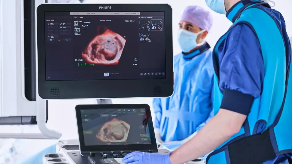
At our clinic, we provide advanced 3D echocardiography imaging to help diagnose heart conditions early and guide effective treatment. This technology allows cardiologists to visualize the heart in real time and make more precise clinical decisions.
Why is 3D Echocardiography Important?
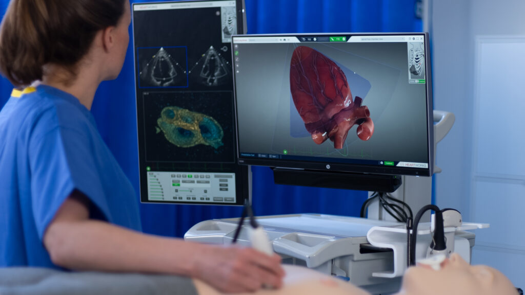
What is 3D Echocardiography?
Using ultrasound waves, the test captures detailed images of the heart valves, chambers, and blood flow, helping cardiologists detect abnormalities and plan treatments more effectively.
How is 3D Echocardiography Performed?
Patient Preparation
No special preparation is usually required. The patient lies comfortably during the test.
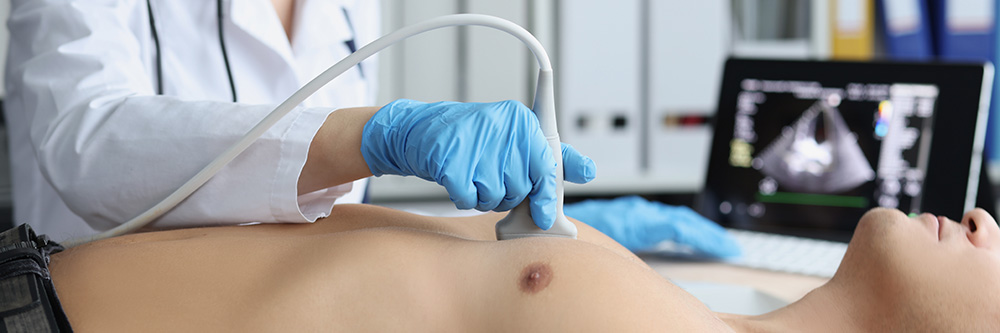
Ultrasound Imaging
A small device called a transducer is placed on the chest, which sends ultrasound waves to create images of the heart.
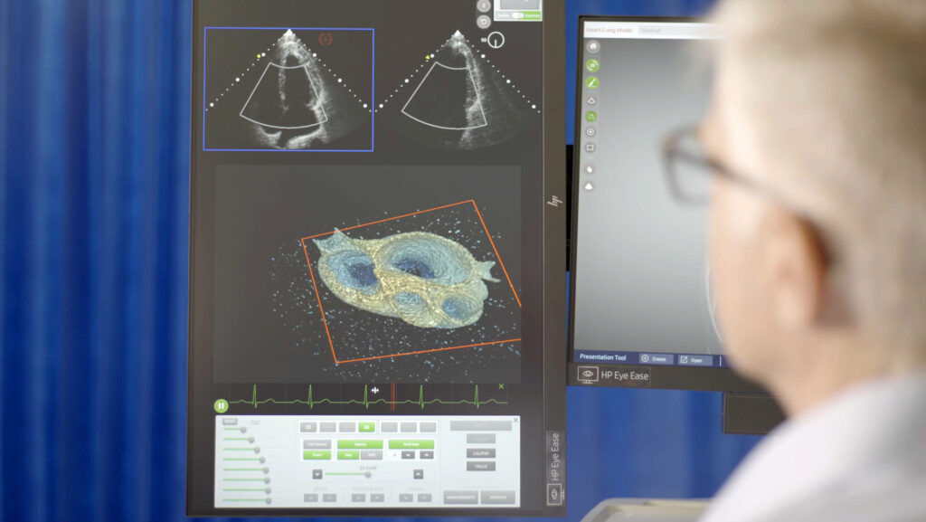
3D Image Generation
The machine captures multiple images and combines them to produce detailed 3D views of the heart.
Procedure Completion
The test typically takes about 20–30 minutes and is completely painless and non-invasive.
Who Should Consider 3D Echocardiography?
3D Echocardiography may be recommended for patients who have:
Heart Valve Disorders: To assess valve leakage, narrowing, or structural abnormalities.
Congenital Heart Defects: To evaluate structural issues present since birth.
Heart Failure: To measure heart function and monitor treatment progress.
Suspected Cardiac Abnormalities: When more detailed imaging is needed beyond standard echocardiography.
Why Choose Us for 3D Echocardiography?
We provide advanced cardiac imaging services using modern technology to ensure accurate diagnosis and effective treatment planning.
Advanced cardiac imaging technology
Experienced cardiology specialists
Accurate evaluation of heart conditions
Patient-friendly and non-invasive diagnostic procedure
Schedule Your Consultation Today
Consult our cardiology team today to ensure early detection and better heart care.